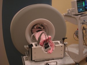
baby mri scan tube
Radio waves cause these. Its a procedure that makes images of your body.

Mri Scans Of A 4 Month Old Infant With Sws A And B Axial Flair Images Download Scientific Diagram
This is the first MRI of a mother kissing her baby.

. For this reason you shouldnt have this test during your first trimester when your babys organs. The aim of the scan is to look for evidence of any blood clot in the lungs called pulmonary embolism PE. It may be used to help diagnose or monitor treatment for a variety of conditions within the chest abdomen and pelvis.
A VQ scan is carried out in two parts. This could be cancerous or a fluid-filled cyst. By combining it with CT or MRI technology healthcare providers are given a more precise portrait of how advanced aggressive or likely a disease may be.
Ultrasound is sound waves with frequencies higher than the upper audible limit of human hearingUltrasound is not different from normal audible sound in its physical properties except that humans cannot hear it. Avoid drinking or eating for a few hours before your scan. They also use it to evaluate the hearts anatomy and function in patients with both heart disease present at birth and heart diseases that.
Most MRI machines are large tube-shaped magnets. It uses a combination of magnetic fields radio waves. The x-ray tube and electronic x-ray detectors are located opposite each other in a ring called a gantry.
A magnetic resonance imaging MRI scanner uses strong magnetic fields to create an image or picture of the prostate and surrounding tissues. Magnetic resonance imaging MRI is a medical imaging technique that uses a magnetic field and computer-generated radio waves to create detailed images of the organs and tissues in your body. Magnetic resonance imaging MRI of the body uses a powerful magnetic field radio waves and a computer to produce detailed pictures of the inside of your body.
The prostate gland is a small soft structure about the size and shape of a walnut which lies deep in the pelvis between the bladder and the penis and in front of the rectum back passage. Your healthcare provider will place a surgical microscope in the ear to get a visual of the auditory canal. Doctors use cardiac MRI to detect or monitor cardiac disease.
This is a type of acoustic evaluation that can help determine the condition of your middle ear and eardrumYour healthcare provider will use various types of air pressure in the ear canal to test the function of. A CT scan is a medical procedure carried out by a machine that creates a 3D image of your body by moving in a circular motion around you. The computer workstation that processes the imaging information is located in a separate control room where the technologist operates the scanner and monitors your examination.
If you are having an ultrasound scan of your digestive system liver or gallbladder you may be asked to. In many ways. Text in the graphic reads.
It can help diagnose problems with soft tissues muscles blood vessels tendons and joints. Basics of CT Scan 1. MRI stands for magnetic resonance imaging.
Eat a low-fibre diet for several days. An ultrasound scan can reveal whether a lump is a tumor. How does the procedure work.
A ventilationperfusion VQ scan is a nuclear medicine scan that uses radioactive material radiopharmaceutical to examine airflow ventilation and blood flow perfusion in the lungs. Cardiac magnetic resonance imaging MRI uses a powerful magnetic field radio waves and a computer to produce detailed pictures of the structures within and around the heart. When you lie inside an MRI machine the magnetic field temporarily realigns water molecules in your body.
This limit varies from person to person and is approximately 20 kilohertz 20000 hertz in healthy young adults. A CT scan is a medical procedure carried out by a machine that creates a 3D image of your body by moving in a circular motion around you. Otoscopy or otomicroscopy.
A PET scan is a sophisticated tool that helps us look beyond the damage caused by a disease to the way in which our body responds to it. Basics of CT Scan 2. Her kiss has caused a chemical reaction in her.
If you are having a pelvic ultrasound scan or are pregnant and are having an ultrasound scan of your unborn baby you may be asked to drink water and not urinate until after your scan. Ultrasound devices operate with frequencies. Menu Healthdirect Free Australian health advice you can count on.
The scan appears to show an adult and a child in a close embrace. Although MRI doesnt seem to harm a growing baby it can raise the temperature inside your body. If you are pregnant the doctor may use body MRI to safely monitor your baby.
Tell your doctor about any.
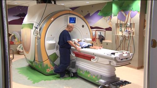
Mri Scans For Babies Youtube

When You Can T Take A Baby To The Mri Machine You Make A Mini Scanner

A Child S Mri With Anesthesia Youtube

Preparing Your Child For An Mri Youtube

Brain Scans Of Premature Babies Reveal Changes That May Raise Risk Of Autism Science The Guardian

Crazy Viral Photo Of Baby In Tube Has A Not So Bizarre Explanation Cafemom Com

Designed With Kids In Mind Child Friendly Mris Stanford Children S Health Bloghealthier Happy Lives Blog
Bringing Mri To Vulnerable Newborns Massdevice
Pediatric Mri